Jan
20
2026
Bastian Asmus
Background
Early iron metallurgy of the first millennium BCE is often described either as technically immature or as a sharp break from Bronze Age practice. Both views are too coarse. Our new study of a bloomery steel chisel from Rocha do Vigio shows a more gradual development (Asmus et al 2026).
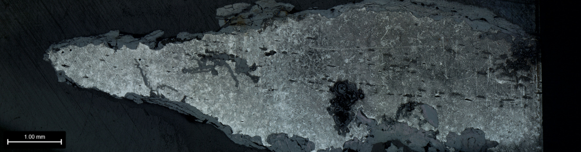
A composite mesoscopic image of the sampled bloomery steel chisel-tip. © 2025 Bastian Asmus
The artefact
The object was made for a bloomery iron and is dated to the ninth century BCE. In an earlier study, we characterised the metal as bloomery steel and determined its carbon content. Quantifying carbon in hardened material is only partly reliable. For this reason, the first study focused on the body of the tool. The cutting edge was not examined at that stage (Araque Gonzalez et al 2023).
Based on a carbon content of about 0.5 wt% C and the early date of the artefact, we later investigated the tip itself. The aim was to assess whether, and to what extent, it had been thermally treated of it was hardened at all.
Bloomery steel: Microstructure and hardness
Metallography of the cutting edge shows a homogeneous and very fine pearlitic to pearlitic-bainitic microstructure. Ferrite is present only in small amounts. Martensite is absent. This points to accelerated cooling, but not to full quenching in the modern sense.
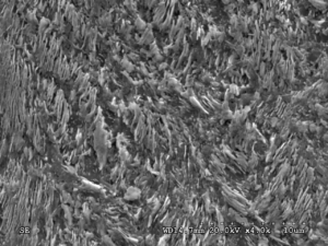
Secondary electron image of the chisel tip close to the cutting edge, showing mostly very fine pearlite, with some upper bainite in between the feathery colonies of the fine pearlite. Image: Asmus.
Vickers microhardness measurements show a moderate hardness gradient between the softer body and the refined tip. The values are consistent with controlled thermal treatment during forging. There is no indication that maximum hardness was the goal.
Alloy chemistry
The bloomery steel is low in manganese, as expected for early iron. Its hardenability therefore differs strongly from that of modern steels. Many commonly used transformation models are based on modern reference compositions. Our results show that these models are only of limited use for early iron artefacts. More accurate transformation data for low-Mn systems are – surprisingly – still lacking.
Production context
Slags from the site confirm local primary iron production. The chisel is part of a regional metallurgical practice. It is not an imported or exceptional object.
Taken together, the evidence points to a deliberate transfer of Bronze Age thermal working strategies to iron. Early iron metallurgy in this case reflects continuity of skill rather than a technological breakthrough.
The full article is available here:
https://doi.org/10.1016/j.jmrt.2026.01.091
References
Araque Gonzalez, Ralph, Bastian Asmus, Pedro Baptista, Rui Mataloto, Pablo Paniego Díaz, Vera Rammelkammer, Alexander Richter, Giuseppe Vintrici, and Rafael Ferreiro Mählmann. ‘Stone-Working and the Earliest Steel in Iberia: Scientific Analyses and Experimental Replications of Final Bronze Age Stelae and Tools’.
Journal of Archaeological Science 152 (April 2023): 105742. doi:
10.1016/j.jas.2023.105742.
Asmus, Bastian, Ralph Araque Gonzalez, Rui Mataloto, Marc Gener-Moret, Pablo Paniego-Díaz, and Pedro Baptista. ‘Negotiating between Iron and Bronze Traditions: The Impact of a Tool – The Chisel from Rocha Do Vigio’.
Journal of Materials Research and Technology 41 (1 March 2026): 1615–29. doi:
10.1016/j.jmrt.2026.01.091.
Comments Off on Bloomery Steel of the Early Iron Age from Iberia. New Article | posted in Analysis, Archaeometallurgy, General, History, Micrograph, Science, slag
Dec
17
2014
Bastian Asmus

 ..for any microscope you might happen to work with. During your microscopy sessions, did you ever wish for less of the dull work, such as noting meta data, contrast method, sample id, photo no or image width? Well – I did.
I did wish for a long time to have a way that my microscope and my camera would speak to each other whenever I change objectives. I am working with Zeiss Universal microscope, mostly with reflected polarising light, i.e. there is no objective revolver. I have to change the objectives individually, which of course, all has to do with the ability to centre the objective for certain steps in polarising microscopy.
To make a long story short: the old days where I have to sit there with a notepad and have to write down all these dull informations are over! From now on my camera, or rather my computer registers any change of my microscope objective and adds this information to my micrographs automatically.
Continue reading
no comments | tags: archaeology, archaeometallurgy, How to, linux, photograhpy | posted in Image Meta information, Lab work, Micrograph, Photography, remote capture, tethered shooting
Jan
29
2014
Bastian Asmus

Fig 1: The use of scientific image processing software allows to quantify the area proportion of each phase in optical micrographs. This is a two step process. The original micrograph is converted to a “threshold map” by modifying the colour channels of the source. The resulting black and white image is analysed for their respective area proportions. The count mask is then produced after quantification and may be used to verify which inclusions have been counted.
 I used this method during my PhD thesis to approximate the chemical composition based on a micrograph .
A traditional method for the quantification of an alloying element in another is the estimation of the carbon content of a steel sample. The area of carbon inclusions is estimated by comparison with known standards, or better by measuring them. Area proportions are believed to represent volume proportions and need to be multiplied with the density ρ to calculate wt% proportions. Continue reading
no comments | tags: How to, photograhpy | posted in Analysis, Archaeometallurgy, General, Micrograph, Microscopy, reflected light microscopy, Science